Glaucoma
What is glaucoma?
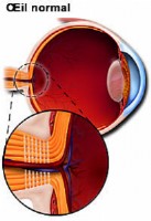
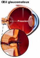
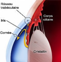
Glaucoma is caused by a number of different eye diseases which in most cases produce increased pressure within the eye. This elevated pressure is caused by the drainage of the fluid produced by the eye that is not enough. This will cause damage to the optic nerve. This damage will become noticeable through the apparition of blind spots in the visual field. If glaucoma is not treated, a gradual vision loss and sometimes even lead to blindness.

Who's at special risk?
Everyone is at risk for glaucoma, however, certain groups are at higher risk than others. It is recommended that people at high risk for glaucoma receive a complete eye exam that includes eye dilatation every one to two years. This is important because early detection, diagnosis and treatment are the only way to prevent vision impairment and blindness.
People at greater risk are:
- People over the age of 45. While glaucoma can develop in younger patients, it occurs more as we get older.
- People who have family history of glaucoma.
- People with abnormally high intraocular pressure.
- People of African descent.
- People who have diabetes, myopia, regular Steroid/Cortisone use.
- People who have suffered from previous eye injury.
Different types of glaucoma
There are a variety of different types of glaucoma. The two main types of glaucoma are primary open angle glaucoma and angle closure glaucoma.
1) Primary Open Angle Glaucoma
This is the most common form of glaucoma. Most people have no symptoms and no early warning signs. If open angle glaucoma is not diagnosed and treated, it can cause a gradual loss of vision. This type of glaucoma develops slowly and sometimes without noticeable sight loss for many years.
2) Angle Closure Glaucoma

This type of glaucoma is much more rare and differs from open angle glaucoma as the eye pressure usually goes up very fast. This happens when the drainage canals get blocked or covered over. Symptoms of angle closure glaucoma may include headaches, eye pain, nausea, seing halo’s around lights at night, and a very blurred vision. Treatment of angle closure glaucoma usually involves surgery to help to unblock the drainage canals so that extra fluid can drain.
3) Normal Tension Glaucoma
In this type of glaucoma, the optic nerve is damaged even though intraocular pressure is not very high. In this case, the optic nerve is probably suffering less from high intraocular pressure than from an insufficient irrigation by the surrounding fessels.
Other forms of secondary glaucoma also exist and may result of an eye injury, inflammation, tumor or in advanced cases of cataract or diabetes. It can also be caused by taking certain drugs as for example steroids.
Diagnosing glaucoma
Early detection, through regular and complete eye exams, is the key to protecting your vision from damage caused by glaucoma. Regular glaucoma check-ups include four main tests.
1) Tonometry
The Tonometer measures the intra-ocular pressure. If the applanation is used , your eye first has to be topically anesthetized. Dr. Vryghem use an instrument that is called air tonometry, a puff of air is sent onto the to take the measurement. Since this instrument does not come in direct contact with your eye, no anesthetic eye drops are required.
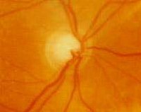
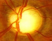
2) Opthalmoscopy
Ophthalmoscopy is used to examine the inside of the eye. The eye doctor will look through the pupil at the optic nerve. Its shape and color can indicate whether or not damage from glaucoma is present and how extensive it is. A dilated retinal exam allows for an even more thorough evaluation of the internal eye.
If the eye pressure is not within the normal range, or if the optic nerve looks unusual, then other special glaucoma tests will be done.
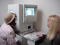
3) Visual field test
In computerized visual field testing you will be asked to look at a fixed point on a special screen and then to press a button each time a light flash is seen. At the end of this test the doctor will receive a printout of your field of vision. This test measures a patient's entire range of vision, including peripheral or side vision.
In patients with glaucoma, typically the side or peripheral vision is lost first, characterized by a narrowing of what is seen outside of center focus.
4) Gonioscopy
Gonioscopy is a painless eye test that checks if the angle where the iris meets the cornea is open or closed, showing if either open angle or closed angle glaucoma is present. This exam is carried out by means of a contact lens placed on the patient’s eye.
5) Optical coherence tomography (OCT)
This is a recently developped method which allows to picture the retina. This technique is far superior to the echography and is mostly used to study macular affections. The OCT instrument utilizes a technique called optical coherence tomography which creates images by use a special beams of light. The OCT machine can create a contour map of the optic nerve , optic cup and measure the retinal nerve fiber thickness. This technique is far more occurate than echography.
Recent discoveries about the cornea are showing that corneal thickness is an important factor in accurately diagnosing eye pressure. Corneal thickness can mask an accurate reading of eye pressure. Actual intraocular eye pressure may be underestimated in patients with thinner corneal thickness, and overestimated in patients with thicker corneal thickness.
More about glaucoma
If you want more information, or would like to make an appointment:
Contact us or call 0032 (0)2 741 69 99