How do the eyes work?
The various parts of the eye each perform a different task in converting light into images.
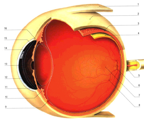
1.The eye muscles are on one side attached to the back of the orbita and on the other to the outer part of the eyeball. The four right muscles attached to the upper, lower and two lateral sides of the eye control its upper, lower and lateral movement. Two oblique muscles control the oblique upper and lower movement.
2.The sclera is the outer layer of the eye. It is the solid, white and opaque tissue to which the muscles adhere. It consists of connective tissue, gives solidity to the eye and protects its inner tissues. It contains the greater part of the eye and merges with the transparent cornea at the front of the eye.
3.The choroidea covers the sclera inside the eyeball. It is a dense maze of small blood-vessels which, amongst other things, feed the cones and rods of the retina. At the place where the sclera merges into the cornea the choroidea merges into the iris, in the middle of which is the pupil.
4.The retina is a wafer-thin, light-sensitive tissue covering the inner part of the eye. It stretches from the place where the optic nerve enters the eyeball to where the choroidea merges with the iris. The retina consists of several layers. The outer or pigment layer is dark in colour due to its pigments. Under that layer is a layer of nerve cells or photoreceptors of which there are two kinds the cones – approximately six million in number – need light to function properly and are able to detect details and colours, the rods need little light and perceive vague shapes in the dark. The retina receives light, the cones and rods transform it into nerve stimuli that are conveyed along the optic nerve to the brain. This is where the images are finally interpreted.
5.The optic nerve carries the stimuli generated by the retina towards the brain, where the image is interpreted. The optic nerve contains more than a million nerve fibres. Each nerve fibre can convey several signals to the brain at the same time. More than fifty per cent of all nerve stimuli received by the brain come from the eyes. The place where the optic nerve leaves the eyeball is called the papilla.
6.The blind spot is about one and a half millimetres in diameter and corresponds to the place where the optic nerve leaves the eyeball. Here there is no retina, so neither cones nor rods are available to produce nerve stimuli and thus light that falls on that spot is not perceived: this is why we call it the blind spot.
7.The macula lies in the centre of the retina, close to the papilla. It is very small but contains a high concentration of cones and is accordingly the most sensitive part of the retina. By means of it we are able to perceive minute details. De gele vlek stelt ons in staat kleine details duidelijk waar toenemen. The further we move away from the macula towards the periphery of the retina the number of cones gradually decreases.
8.The blood vessels bring nutrition to the eye.
9.The vitreous body or corpus vitreum is a clear gelatinous substance, surrounded by a thin membrane, that fills the inner part of the eye and pushes the inner layers of the eye against its outer wall.
10.The corpus ciliare is a thickening where the choroidea merges into the iris. It consists of a ring of muscles which hold the lens in place.
11.The posterior chamber of the eye is where aqueous humor, a clear fluid containing nutritional substances for the cornea, the lens and the vitreous body, is produced. This space lies behind the iris at the place where the lens hangs.
12.The anterior chamber is the space between cornea and iris. Because aqueous humor is constantly produced and evacuated the pressure in the eyeball remains constant.
13.The pupil is the dark opening in the middle of the iris. Light enters the eye through the pupil. In bright light, as in sunlight, the pupil narrows to protect the retina. In lesser light, as in a dark room, the pupil widens to allow more light to enter the eye.
14.The iris is the coloured part of the eye lying behind the cornea. The opening in the middle of it is called the pupil. The pupil widens and narrows, thereby determining how much light enters the eye. If too much light comes in the circular muscle of the iris contracts, as a reflex, and the pupil narrows. With less light the pupil narrows. The colour of the iris is determined by the number of pigment cells. If there are many of these, the iris is brown; if there are fewer, the iris is blue or grey.
15.The cornea is is the transparent, clear tissue at the front of the eye. Light enters the eye through the cornea’s wide surface and is directed towards the pupil. Two thirds of the refractive power of the eye, which directs the light rays towards the retina, is due to the cornea. It contains many nerve ends and so is very sensitive. To avoid drying up, tears are spread by blinking over the surface of the cornea.
16.The crystalline lens is the clear lens which hangs behind the pupil. Together with the cornea it ensures that the light rays entering the eye are concentrated and directed so as to fall precisely on the retina. This process is called refraction. The flexibility of the lens enables it to become flatter or steeper, by means of an internal muscle, depending on the distance from the object viewed. This process is called accommodation. In this way an object, whether near or far away, can be seen as a sharp image. The image on the retina is an inverted picture of the object whose light rays are received by the eye.
If you want more information, or would like to make an appointment:
Contact us or call 0032 (0)2 741 69 99